Full Text
Introduction
Sickle cell disease (SCD) is an inherited disorder caused by the single point mutation due to the replacement of glutamic acid to valine at the sixth codon of β-chain of globins (β6Glu→Val) [1], which is an alternation of single nucleotide base from thymine to adenine (GTG → GAG) the molecular change is responsible for the alternation in the properties of the haemoglobin tetramer, with a tendency to polymerize in the deoxygenated state altering normal, flexible, biconcave shaped red blood cells (RBCs) in to stiff, rigid, sickle cell RBCs [2]. These changes is related to fundamental pathophysiology, vaso-occlusion, tissue ischemia, sickle shaped erythrocytes, leucocytes, platelets and endothelial cells [2].
Hepatic dysfunction is usually known problem of SCD due to multiple factors such as intrahepatic sinusoidal sickling and transfusion related hepatic infections [3]. The sickle cell disease with hepatic dysfunction shows the abnormal variations in liver function tests including serum aminotransferases, bilirubin and alkaline phosphatase [4].
Our study focusing on the clinical investigation of serum aminotransferase [serum glutamic oxaloacetic transaminase (SGOT) and serum glutamic pyruvic transaminase (SGPT)] enzymes.
Material and methods
Place and ethical clearance of the study
This study was conducted from 2019 to 2020 in the Department of Biochemistry, People’s College of Medical Sciences and Research Centre (PCMS&RC) and Centre for Scientific Research and Development Department (CSRD), People’s University, Bhopal, India. The study protocol and ethical clearance had been approved by the Research Advisory Committee and Institutional Ethical Committee.
Sample size estimation
The sample size estimation had been carried out and confirmed by the expert statistician. The estimated value was based on the prevalence of sickle cell disease in Madhya Pradesh. The prevalence was found in between the range of 15% to 30% [5]. It was carried out by using the formula: N= 4PQ/L² . The estimated sample size was found to be 110.68. Hence 111 SCD samples of cases or more samples are necessary to meet the desired statistical constraints. So 111 healthy subjects were enrolled in the present study with their consent forms.
Study design
This is a hospital based, case control, prospective study. The blood samples were collected from the People’s Hospital and Civil Hospital Bairagarh, Bhopal, Madhya Pradesh.
Study criteria
Inclusion criteria for sickle cell disease subjects: (a) The subject should have diagnosed with SCD with other sickle cell related complications alongside acute sickle pain, including not limited to acute chest syndrome, renal dysfunction, liver dysfunction stroke, vaso-occlusive event, and priapism have been included in the study, (b) Patients more than 5 and less than 70 years of age was included in the study. All the selected and enrolled patients had been confirmed with their specific characters with a single blood transfusion of 3-4 months before the blood drawn. The patients with hospitalization were included in the study and were might be or might not be under the continuous antibiotics therapy, hydroxyurea (HU) treatment. The patients under hydroxyurea treatment advised to stop hydroxyurea for 15-20 days, and after that the blood to be collected for the research study.
Exclusion criteria for sickle cell disease subjects: (a) Sickle cell subjects less than 5 and more than 70 years was excluded from the study, (b) Individuals with other hemoglobinopathy was also be excluded from the study, (c) SCD patients under chemotherapy treatment were excluded from the study, (d) For the safety precautions in handling the blood samples, patients with HIV, hepatitis B, and hepatitis C were excluded from the study.
Source of normal healthy subjects
The control group consist 111 healthy normal individuals, healthy persons were comprised of departmental staff, medical students and relatives who were healthy and accompany their Outpatient Department (OPD) or Inpatient Department (IPD), and their health condition was detected.
Conformation tests for SCD
The screening tests were carried out with SCD patients for the confirmation of sickle cell RBCs. (a) Sickling test with 2% meta-bisulfite: It is the principle of the sickling test, was based on microscopically observation of sickling of red blood cells when exposed to low oxygen tension. (b) Solubility test with 0.02% sodium dithionate: It is the principle of solubility method, based on turbidity created when Hb-S is mixed with sodium dithionate. (c) Peripheral blood film method: Thin blood films, stained with giemsa stain were examined by light microscopy (×100). (d) Hb electrophoresis: The cellulose acetate membrane Hb electrophoresis method was used to determine the presence of Hb-S in the sample.
Serum SGOT and SGPT were estimated by Reitman & Frankel’s method [6] and were diagnosed by using the Trans Asia diagnostic kits (Manufactured by Transasia Bio-Medicals Ltd., B-11, and OIDC, Ringanwada, Daman-396210, India) in collaboration with ERBA diagnostics Mannheim HmbH Mallaustr, 69-73, D-68291, Mannheim/ Germany.
Statistical analysis
The statistical calculation was done by using the SPSS software version-24. In this study we observed that, SGOT and SGPT were found highly significant in sickle cell disease cases as compared to the controls and the Pearson’s coefficient correlation shows us positive correlation with each other. The P ˂ 0.000 was considered to be statistically highly significant.
Results
Changes in transaminases of patients with sickle cell disease versus controls, mean SGOT and SGPT levels in the overall population of 111 subjects were 69.98 ± 69.31 and 25.17± 5.25 IU/L, respectively. The highest SGOT/AST measurement was 139.29 IU/L, and the highest for SGPT was 30.42IU/L. However, for 111 non sickling subjects, the mean SGOT and SGPT levels were the 65.28 ± 60.07& 22.72 ± 5.47 IU/L, mean SGPT and SGOT levels for the first determination were 125.35 and 28.19 IU/L, respectively. Mean SGOT and SGPT levels with sickle cell disease were higher than in their control counterparts, while the mean SGOT, was higher than SGPT (Table 1), but these differences were highly significant (P < 0.000). However, the mean SGOT levels of both male and female subjects with sickle cell disease were significantly higher than for controls (P < 0.000) for both genders. None of the differences in SGPT levels were significant. All the differences in SGOT levels between the SCD and normal subjects were highly significant (P < 0.000). In 95.8% of the subjects with sickle cell disease, the SGOT levels were increased than SGPT, but our results did not suggest overt malfunctioning of the liver and heart in the majority of subjects. In the present study, the mean standard deviations of cases vs. controls were showed significant difference.
There was the highly significant difference observed in between patients and controls in SGOT and SGPT variables, since the p ˂ 0.000 (Table 1).
Table 1: Distribution of SGOT and SGPT variables among the patients and controls.
|
Variables
|
Group
|
N
|
Mean
|
SD
|
‘t’ value
|
‘p’ value
|
Significance level
|
|
SGOT IU/lit.
|
Patients
|
111
|
69.98
|
69.31
|
6.79
|
˂ 0.000
|
Highly significant
|
|
Control
|
111
|
25.17
|
5.25
|
|
SGPT IU/lit.
|
Patients
|
111
|
65.28
|
60.07
|
7.43
|
˂ 0.000
|
Highly significant
|
|
Control
|
111
|
25.17
|
5.25
|
Note: p˂ 0.0001= highly significant and p˂ 0.005 significant.
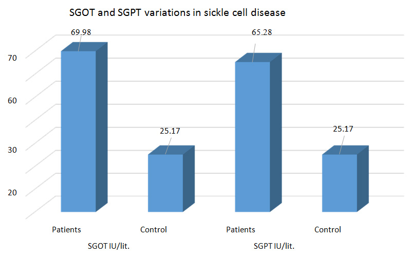
The graph designating that there was severely increased level of SGOT (69.98IU/L) and SGPT (65.28IU/L) found in the sickle cell disease patients as compared to the control group. The Mean values of the controls were with in normal limit with 25.17IU/L for both SGOT & SGPT (Figure 1).

Figure 1: Distribution of Sr. SGOT and SGPT level variations in SCD.
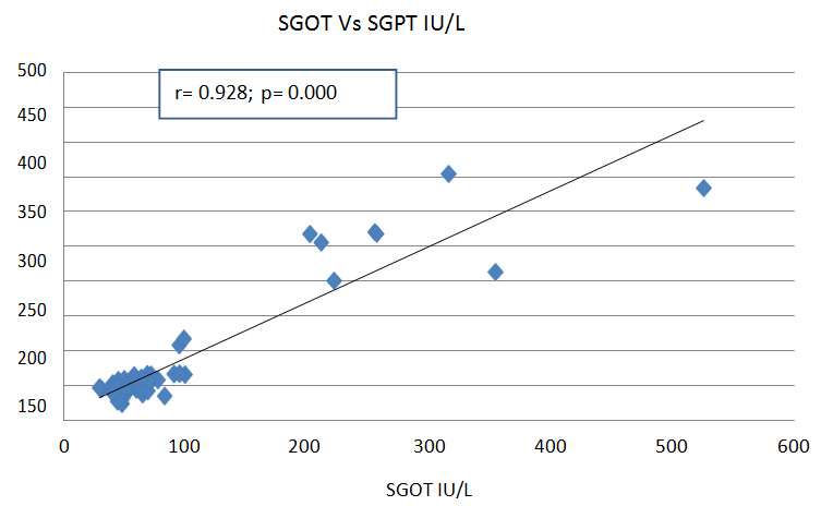
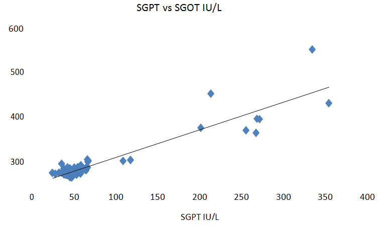
The figure 2a,b shows a designating Pearson’s coefficient correlation analyses were carried out to find the association between the SGOT vs SGPT and vice verso. In the simple Pearson’s coefficient correlation analyses, the SGOT vs SGPT and SGPT vs SGOT was the dependent variable (Figure 2). There was a positive correlation between SGOT vs SGPT levels. Thus, the most highly significant association was found for the combination of SGOT vs SGPT. A striking feature is that it was in the crisis states that the relationship assumed high statistical significance.

Figure 2a: Pearson’s coefficient correlation.

Figure 2b: Pearson’s coefficient correlation.
Discussion
The activity of SGOT and SGPT has widely used liver function tests to assess the liver conditions; both enzymes are very sensitive and reliable for the assessment of LFT. These enzymes was increased in SCD due to the organ damage property due to sickling mechanism which causes to liver dysfunction [7].
In our findings, the liver enzymes SGOT and SGPT were found increased and showed significantly high in SCD cases but in the controls were found within the normal levels.
An increase in SGOT and SGPT is due to the rapid sickling of RBCs in sickle cell disease; the transamination was not complete and were not utilized for the conversion of keto acids which leads to increase the levels of SGOT and SGPT [8].
Our outcomes are correlated with Richard et al study [9], they stated that the significant alterations in the levels of liver enzymes and other parameters were also seen in all the patients of SCD as compared to controls (p<0.0001), were the LFT parameters were found significantly (p<0.0001) elevated in cases. But in the steady state of sickle cell disease, Richard et al [9] studied the same variables in which he reported that, there were no significant alterations in the levels of liver function enzymes, that could be due to the steady state of the patients where sickling and hemolysis was not rapid [8].
Mahera et al and Gardner et al concluded in their studies that, there was a significant increase in LFT parameters which were similar in both results [10, 11]. Gardner K et al also studied a milder increase of LFT which were also observed in the SCD cases [11].
We have also studied the enzyme activities by using Pearson’s correlation coefficient which showed very significant positive correlation, which is supported by the different studies that is discussed here below:
Akuyam et al found that there were statistically highly significant increased levels of Total bilirubin, Sr. AST, Sr. ALT and ALP in sickle cell disease [8]. Gardner et al and Brody et al stated through the observations of his study that, the previous elevations of the Sr. AST (94.4%), ALT (2.8%), were found more in SCD patients [11, 12]. Norris et al reported that, the acute sickling process selectively affects to the liver in 10% of the patients, causing the liver crisis with abdominal pain, nausea, fever, jaundice and were found the elevated levels of SGOT and SGPT in SCD [13].
Similarly, Johnson et al, Sheehy and Stephan et al were stated that, the liver enzyme transaminases were found significantly increased in sickle cell hepatopathy, which was due to the rapid sickling and hemolysis of the red blood cells [14-16].
Schubert et al and scientists studied that the serum alkaline phosphatase level was also markedly elevated in the sickle cell disease, but the ratio of serum aspartate amino transferease and serum alanine amino transferease enzymes were not altered significantly [4, 13-15].
In our study graph-3 indicates that there is the significant positive Pearson’s coefficient correlation between SGPT and SGOT. Where r= 0.929; P=0.000.
They also explained that, Sr. AST enzyme was mostly found elevated excessively due to an increased rate of hemolysis of RBCs in sickle cell disease and significantly increase in levels of serum bilirubin was also reported due to the ongoing and increased rate of hemolysis; in intrahepatic cholestasis and renal impairments encountered in sickle cell hepatopathy in comparison with the other remaining all diseases [4, 13-15].
Thus, the study could be concluded that, an increase in SGOT, SGPT, might be due to the rapid sickling of RBCs in sickle cell disease.
Conclusion
It is concluded that SGOT and SGPT enzymes were significantly increased in sickle cell disease, so it would be another laboratory parameters for the diagnosis of SCD. This study has shown that, in patients with sickle cell disease has a haemolytic tendency attributable to mutant haemoglobin S in red cells, there is generally higher elevation of SGOT relative to SGPT, leading to good Pearson’s correlation of coefficient. This finding is at variance with the generally held view that, in patients with acute and chronic liver disease of various origins, the SGOT, SGPT provides useful clinical information regarding both the cause and severity of liver disease. We therefore propose use of the SGOT, SGPT in sickle cell disease as a haemolytic index, and using simple and Pearson’s correlation analyses, between the SGOT & SGPT showed highly significant changes.
Acknowledgement
The staff of Departmental of Biochemistry for their timely important advice, help and encouragement.
Conflicts of interest
Authors declare no conflicts of interest.
References
[1] Rees GP, Williams TN, Gladwin MT. Sickle cell disease. Lancet. 2010; 376(9757):2018–2031.
[2] Ballas SK. Sickle cell anemia: progress in pathogenesis and treatment. Drugs. 2002; 62(8):1143–1172.
[3] Kakarala S, Lindberg M. Safety of liver biopsy in acute sickle hepatic crisis. Conn Med. 2004; 68(5):277–279.
[4] Schubert TT. Hepatobiliary system in sickle cell disease. Gastroenterology. 1986; 90(6):2013–2021.
[5] Gupta VK, Singh N, Masram SW, Patil SKB. Status of some hematological and antioxidant parameters in SCA patients of Chhattisgarh region. J Medl Sci Clinic Res. 2015; 7:3.
[6] Reitman S. Frankel S. A Colorimetric method for the determination of serum glutamic oxalacetic and glutamic pyruvic transaminases. Amer. J. Olin. Path. 1957; 28(1):56–63.
[7] Tiez NW. (Ed) Text book of Clinical Chemistry. 1986; pp.1388.
[8] Akuyam AS, Bamidele AS, Aminu SM, Aliyu IS, Muktar HM, et al. Liver function tests profile of sickle cell anemia patients in steady state of health: Zaria experience. Borno Med J. 2007; 4:1–6.
[9] Richard S, Billett HH. Liver function tests in sickle cell disease. Clin Lab Haematol. 2002; 24(1):21–27.
[10] Mahera MM, Mansour AH. Study of Chronic Hepatopathy in Patients with Sickle Cell Disease. Gastroenterology Research. 2009; 2(6):338–343.
[11] Gardner K, Suddle A, Kane P, O’Grady J, Heaton N, et al. How we treat sickle hepatopathy and liver transplantation in adults. Blood. 2014; 123(15):2302–2307.
[12] Brody JI, Ryan WN, Haidar MA. Serum Alkaline phosphatase isoenzymes in sickle cell anemia. JAMA. 1975; 232(7):738–741.
[13] Norris WE. Acute hepatic sequestration in sickle cell disease. J Natl Med Assoc. 2004; 96(9):1235–1239.
[14] Johnson C, Omata M, Tong M, Simmons J, Weiner J, et al. Liver involvement in sickle cell disease. Medicine.1985; 64(5):349–356.
[15] Sheehy TW. Sickle cell hepatopathy. South Med J. 1977; 70(5):533–538.
[16] Stephan IJ, Merpit–Gonon E, Richard O, Raynaud-Ravni C, Freycon F. Fulminant live failure in a 12 year old girl with sickle cell anemia: favourable outcome after exchange transfusions. Eur J Pediatr. 1995; 154(6):469–471.